Page 723 - My FlipBook
P. 723
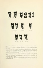
Q Q 99 99
Fig. 432. Fin. 43.S. Fig. 435.
tiv 1 ^
Fid. 436. Fu;. 43 Fig. 430.
Fin. 440. I'"h;. 441. Fig. 442.
Figs. 432, 433. Mesio-distal sections of lower first molars, exposing the pulp chamber and root
canals.
Fig. 434. A bucco-lingual section through the mesial root of a lower first molar, showing the
Not very infrequently these end
pulp chanilier and the two canals in the mesial root. in a common
npical foramen.
Fig. 435. Cross-sections through the central part of the pulp chambers of two lower molar teeth
in the figures one above the other on the left. The second cut is just below the pulp chamber in each.
The third cut is about the mid-length of the roots. Tn the upper series, the mesial root has one broad
canal. In the lower series this is divided into two very small canals, widely separated.
Fig. 436. A mesio-distal section of a lower second molar with a single root, showing the pulp
chamber and a mesial and distal root canal.
Fig. 437. A bucco-lingual section of a lower second molar with one root and one large pulp canal.
Fig. 438. A mesio-distal section of a lower second molar with two roots, showing the root
canals.
Fig. 439. A bucco-lingual section of the crown and mesial root of a lower second molar, with
two root canals which join in the apical third of their length and again separate, ending in separate
apical foramina. This is unusual.
Figs. 440, 441, 442. Mesio-distal sections of lower third molars, showing the forms of the pulp
chambers and root canals. Figure 441 shows the single root with one large canal. This is not very
uncommon in these teeth.