Page 721 - My FlipBook
P. 721
DlkftOO»
Fig. 422. Fig. 423.
Sty
Pig. 424. Fig. 425. Fig. 4-2G. Fig. 427.
(9 R f
Fig. 431). FlG. 4al.
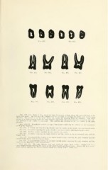
Figs. 422. 423. Each of these represent three horizontal sections across tlie neck and root of an
upper first molar. The first in each, reading from left to riglit, is through the central part of the pulp
chamber. The second is at the point where the canals are dividing from the chamber. The third
section IS a little rootwise from the pulp chamber and shows the molar triangle, formed by the
rela-
tive position of the canals, to advantage. This exhibits the relation of the root canals to the duId
chamber. ^ '
,FlGS. 424-427. Lengthwise sections of upper first molars exhibiting the relation of the root canals
to the pulp chambers.
Flo. 424. A section exposing the pulp chamber and the canals in the mesio- and disto-buccal roots.
Fia. 425. A section exposing the pulp chamber and mesio-buccal and lingual pulp canals.
Fig. 426. Another section exposing the two buccal root canals.
Fio. 427. A section exposing the pulp chamber and the canals in the disto-buccal root and the
lingual root.
Fig. 428. A perpendicular section of an upper second molar, exposing the pulp chamber and the
canals in the mesio- and disto-buccal roots.
Fig. 429. A peiijendicular section of an upper second molar, exposing the pulp chamber and the
canals in the disto-buccal and lingual roots.
Figs. 430, 431. The pulp chamber and root canals in upper third molars. Figure 430 is a
bucco-lingual section showing the canals in the mesio-buccal and lingual roots.
Figure 431 is a mesio-
'
distal section showing the divided buccal canals in a tooth with a single root