Page 298 - My FlipBook
P. 298
134 THE TECHNICAL PROCEDURES IN FILLING TEETH.
pressure on the paper. When the cement hecomes hard, the
paper may be pulled away.
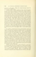
Another matter briefly mentioned above, that frequently
becomes important, is shown in the split cavity. Figures 138-141.
In this case, as shown in Figures 138, 139, of a molar tooth split
mesio-distally, there are two decays, one in the central pit and
one in the distal pit, both extending widely along the dento-
enamel junction, almost completely undermining the enamel of
the occlusal surface in the mesio-distal direction. This has gone
so far that only the marginal ridges on the mesial and distal
can be retained. If the dentin walls were cut parallel with the
long axis of the tooth, the strength of the dentin remaining
would be insufficient. In such cases, the mesial and distal walls,
one or both, should be so sloped as to retain sufficient strength
of dentin, as shown in Figures 140, 141. To the buccal and to the
lingual of the cavity, there is abundant material and a sufficient
part of these walls may be made parallel to render the filling per-
fectly secure. This is especially true in cases in which the depth
in relation to the pulp, the patient being of mature age, allows
the full squaring out of the pulpal wall, as represented in this
case.
It should be noticed also that the disto-lingual groove in
Figure 140, and the buccal groove in Figure 141, were each made
with a cut which only entered the dentin a little, not cutting this
part to the depth of the principal cavity. Deep cutting in these
position weakens the tooth without accomplishing any mechan-
ical or protective advantage. Cuts following out grooves in
order to find points to make a good tuiish, should always be deep
enough for independent anchorage of the ends farthest out from
the cavity. In these illustrations the enamel cap has been made
sufficiently prominent for the dento-enamel junction to be fol-
lowed in all of the parts. It should be particularly noted that this
line rises as the enamel is cut back onto the slopes of the cusps,
apparently deepening the cavity. The size and form of the pulp
chamber in this case represents greater age of the patient than
that in the previous series of illustrations, gi\"ing more freedom
in squaring out the pulpal wall.
Figures 142-144 represent a tooth with decay, cavity pre-
pared, and filling placed, in a similar case to that sliown in Fig-
ures 138-141. The excavation of such cavities as these is carried
out in a manner practically the same in its details as that
represented in Figures 128-137. There is considerably more
chipping of enamel in uncovering the areas of decay, and some-