Page 259 - My FlipBook
P. 259
Fio. 110.
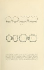
Fms. llfi. 117. A diagrammatic representation of caries «f the buccal surfaces. Figure IIG. anJ
of cross sections of the crowns. Figure 117, of the lower first and second bicuspids and first and
second molars, showing the location of caries and tendency to spread in a direction around the
cr<'WDs of the teeth, following the free margin of the gingi^Te. In Figure 116, the dotted line repre-
sents the gingival line, or line of the attachment of the gimi tissue to the teeth. The continuous
dark line reprt-stnts the line of the free margin of the gingi^ie, which arches toward the occlusal
in passing between the teeth. The double line represents a saw cut dividing the crowns through the
decayed areas of the enamel. Figure 117 represents the decayed areas of enamel exposed by cutting
away the crowns. The portions darkeiieii re]>resent the parts of the enamel mast liable to caries,
while the areas left white at the mesio- and dislo-buccal angles of the teeth represent areas that are
almost always immune to caries.