Page 257 - My FlipBook
P. 257
Kk;. hi.
Fin. 114. A diagrainniatic representation of a cross section of a lower molar, showing- beginnini?
decays in the proximal surfaces and in the buccal surface. In these the directions of the penetra-
tion are marked by arrows. Notice that these are broader than in Figure 112, for the reason that
the greater spreading of decay of the enamel is in a direction around the crown of the tooth.
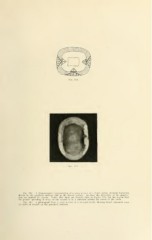
Fig. 11;i. .V photograph from a cross section of a bicuspid tooth, showing broad whitened areas
iif caries of enamel on the proximal sm'faces.