Page 275 - My FlipBook
P. 275
DIAGNOSIS AND PROGNOSIS 243
this will be noted the lamina dura, exhibited in the form of
a radiopaque line. The trabecula of bone at the apex and
outside of this line should appear homogeneous with the
surrounding bone.
In some instances the apical space surrounding teeth
recently infected or infected with micro-organisms of low
virulence may appear perfectly healthy in the X-ray film,
while teeth suffering from the effects of traumatic occlusion,
or chronic inflammatory disturbances of the apical tissues
may exhibit an apparent rarifying process in the surrounding
bone, simulating infection. Consequently, it should be
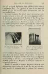
Fig. 278.—Small granuloma on the Fig. 279.—Granuloma on lower
cuspid. Canal unfilled. anterior teeth. Teeth badly af-
fected with pyorrhea.
remembered that the appearance of a radiolucent area at the
apex does not always signify the presence of infection.
Many errors in diagnosis are thus made from an imperfect
understanding of this fact. The radiograph, is then, not an
infallible guide in the diagnosis of infective conditions of
the periapical tissues.
When it is decided to attempt the preservation of teeth
affected with septic conditions at the root end, the responsi-
bility should be assumed alike by patient and operator.