Page 166 - My FlipBook
P. 166
134 PREPARATION OF CAVITIES
enamel from the buccal side with a chisel, gradually shifting
more and more to the occlusal and continuing until the ridge
is sufficiently removed.
After removal of the marginal ridge, extend the cavity
in the form of a step, by means of the fissure and inverted
cone burs and hatchet excavators, the full length of the
occlusal groove (bicuspids), or to include the entire central
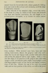
Fig. 165. Fig. 166. Fig. 167.
Fig. 165.—Step form in a disto-incisal cavity on an upper cuspid.
Fig. 166.—Labial view of a mesio-incisal cavity on an upper cuspid showing flat
gingival wall.
Fig. 167.—The incisal step form applied to a distal cavity on an upper cuspid.
The labial enamel has been preserved.
fossa (molars). With inverted cone burs, hoes and side
cutting instruments, flatten the gingival wall (the seat),
make the buccal and lingual walls parallel with each other
and joining the gingival wall at right angles. Make a defi-
nite line angle at the junction of the gingival and axial walls
(gingivo-axial) and definite point angles (bucco-gingivo-axial
and linguo-gingivo-axial) at the buccal and lingual termina-
tions of this line. Flatten the top of the step, the pulpal
wall, and cut that portion of the cavity, as well as the proxi-
I
enamel from the buccal side with a chisel, gradually shifting
more and more to the occlusal and continuing until the ridge
is sufficiently removed.
After removal of the marginal ridge, extend the cavity
in the form of a step, by means of the fissure and inverted
cone burs and hatchet excavators, the full length of the
occlusal groove (bicuspids), or to include the entire central
Fig. 165. Fig. 166. Fig. 167.
Fig. 165.—Step form in a disto-incisal cavity on an upper cuspid.
Fig. 166.—Labial view of a mesio-incisal cavity on an upper cuspid showing flat
gingival wall.
Fig. 167.—The incisal step form applied to a distal cavity on an upper cuspid.
The labial enamel has been preserved.
fossa (molars). With inverted cone burs, hoes and side
cutting instruments, flatten the gingival wall (the seat),
make the buccal and lingual walls parallel with each other
and joining the gingival wall at right angles. Make a defi-
nite line angle at the junction of the gingival and axial walls
(gingivo-axial) and definite point angles (bucco-gingivo-axial
and linguo-gingivo-axial) at the buccal and lingual termina-
tions of this line. Flatten the top of the step, the pulpal
wall, and cut that portion of the cavity, as well as the proxi-
I