Page 94 - My FlipBook
P. 94
32 PATHOLOGY OF THE HARD TISSUES OF THE TEETH.
cut them out and make fillings. There is no reason for doing
this unless softening has occurred, or in other words, unless
decay has actually begun. Of course in that case the filling
is the proper procedure, but not otherwise.
Other Deformities op the Ekamel.
In examining the teeth of many people, other deformities
of the enamel than atrophy and the pits above described are
occasionally met. It is desirable that these be distinguished
and separated from typical atrophy. Generally, this is easy
if we keep the typical forms of atrophy in mind. Atrophy, due
to suspension of nutrition, affects certain parts of certain teeth,
following the lines of contemporaneous development, and when
this is seen in any one tooth, certain parts of certain other teeth
are marked and all others have escaped. Therefore, if we find
a ease in which a certain part of each tooth of the whole set is
pitted and rough, or grooved, the injury or deformity not fol-
lowing the lines of contemporaneous development, we have
found another condition entirely and should separate it sharply
from the form of atrophy above described. How many such
cases have been confounded with atrophy in the past that have
no histological or causal relation to it can not now be told. That
some have, there is no longer room for doubt. I have collected
a number of such eases. I shall describe only some of the most
notable cases coming under my observation. I shall not try to
group these into special kinds of deformities, for the reason that
I have not studied a sufficient number to arrive at general con-
clusions.
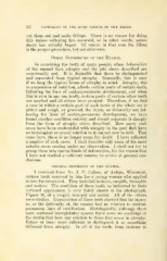
general deformity of the enamel.
I received from Dr. J. E. Callow, of Antigo, Wisconsin,
sixteen teeth removed by him for a young woman who applied
to him for treatment. They included incisors, cuspids, bicuspids
and molars. The condition of these teeth, as indicated by their
outward appearance, is very fairly shown in the photograph,
Figure 36, of a cuspid, bicuspid and molar. All of the others
were similar. Examination of these teeth showed that the injury
to, or the deformity of, the enamel had no relation to contem-
poraneous lines of calcification. Histologically, although there
were scattered interglobular spaces, there were no markings in
the dentin that bore any relation to those that occur in atrophy.
Either of these were sufficient to distinguish it as something
different from atrophy. In all of the teeth, from incisors to