Page 83 - My FlipBook
P. 83
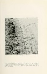
Fia. 32.
Fig. 32. A photomicrograph with a much higher power from another tooth with a similar atrophy
to that shown in Figure 31, in which the section was cut horizontally or crosswise. The section through
the interglobular spaces is, therefore, through that portion of the line showing in Figure 31, which
dips toward the gingival at the buccal and lingual portions of the section. e. Enamel. d. Dentin.
8. s. Line of interglobular spaces appearing as irregular dark blotches connecting the one with the
titbiM" in every direction.