Page 42 - My FlipBook
P. 42
14 PATHOLOGY OF THE HAED TISSUES OF THE TEETH.
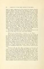
chart, or index, indicating by lines across the incisors, cuspids
and first molars, the positions the grooves across the teeth
assume because of disease occurring at different ages of the
child. Figure 9. These lines have been varied a little from the
true contemporaneous calcification lines to suit better the appar-
ent positions upon teeth that are shortened by severe atrophy.
This chart will point out the age at which any injury occurred
as well, perhaps, as it can be done in a chart of this character,
which, of course, is founded upon averages. Pretty wide varia-
tions will occur in the time of the calcification of the teeth of
individuals, and also between the several teeth of the same indi-
vidual. There is certainly as much variation as eighteen months
in the time of the beginning of the calcification of the central
incisors, and a greater range possibly with all of the other teeth,
except the first molars. These latter are perhaps the most con-
stant. But these certainly vary from the twenty-fifth week of
uterine life to something near six months after birth.
The chart is intended to give only a general average as to
the time of the illness that has caused these injuries.
HisToiiOGiCAii Chabactees in Atrophy of the Teeth.
In presenting the histological characteristics in atrophy of
the teeth, it may be stated that all of the cases thus far examined
by myself, no matter how different their outward appearance,
present one plan of departure from the normal arrangement
of tissues. The differences are due only to position, the number
of zones of injury and in the details of severitj^. This plan is
inseparably linked with the plan of development of the dental
tissues. There are, however, other forms of deformity of the
enamel in a degree simulating atrophy, which are entirely dif-
ferent in histological characters. Some of these will be noted
later.
Except in the pits that often accompany it, the zones of
injury in atrophy rigidly follow the lines of Retzius. In the dia-
gram, FigTire 10, the lines of Retzius are made especially prom-
inent to recall distinctly their direction on different parts of the
enamel cap of the crown of the tooth. In microscopic observa-
tion these are usually clearly seen in some parts of the enamel
cap, particularly in central labio-lingual sections. They vary,
however, indefinitely in prominence in different sections, and in
different parts of the same section. Generally, they do not show
clearly in all parts of a section, and those who have not studied
them carefully should refresh their memory as to the course of