Page 34 - My FlipBook
P. 34
10 PATHOLOGY OP THE HARD TISSUES OF THE TEETH.
It is not frequent that we see so severe a mark as here shown
so high upon the labial surfaces of the incisors. It seems to be
a general rule that the higher upon the teeth the less marked is
the deformity. Pretty generally, in this position on the cen-
trals, the mark is a shallow groove, more or less pitted, or a row
of pits without a distinct groove. In all of these cases the lower
teeth bear marks similar to those in the upper.
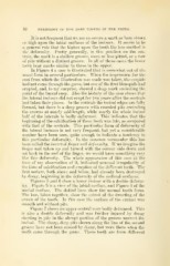
In Figure 4 a case is illustrated that is somewhat out of the
usual form in several particulars. When the impression for the
cast from which the illustration was made was taken, the cuspids
had not come through the gums, but one of the first bicuspids had
erupted, and, to my surprise, showed a deep mark encircling the
point of the buccal cusp. Also the history of the case shows that
the lateral incisors did not erupt for two years after the centrals
had taken their places. In the centrals the incisal edges are fully
formed, but there is a deep groove with rounded pits encircling
the crowns at nearly mid-length, while nearly the whole incisal
half of the laterals is badly deformed. This indicates that the
beginning of the calcification of these teeth was late, as compared
with that of the centrals. This particular form of deformity of
the lateral incisors is not very frequent, but yet a considerable
number have been seen, quite enough to indicate a tendency to
this particular deformity. In the common vernacular this has
been called the inverted finger nail deformity. K we imagine the
finger nail taken up and turned with the convex side down and
set back in the end of the finger, we would have something very
lilce this deformity. The whole appearance of this case at the
time of my observation of it, indicated imusual irregularity of
the time of calcification and eruption of the different teeth. The
first molars, both above and below, had already been destroyed
by decay, beginning in the deformity of the occlusal surfaces.
Figures 5 and 6 show a lower incisor with a double deform-
ity. Figure 5 is a view of the labial surface, and Figure 6 of the
mesial surface. The dotted lines show the normal tooth form.
The two, taken together, show the extent of the dwarfing of the
crown of the tooth. In this case the surface of the enamel was
smooth and without pits.
Figure 7 shows an upper central very badly deformed. This
is also a double deformity and was further injured by decay
starting in pits in the abrupt portion of the groove nearest the
incisal. The sharp, deep pits shown along the line of the second
groove have not been caused by decay, but were there when the
tooth came through the gums. These teeth are from different