Page 293 - My FlipBook
P. 293
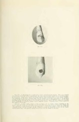
Fig. 135.
Fig. 134. A photograph of a cuspid with a decay across its labial surface. There is a decayed
area nmuins across the surface mcsio-distally that has penetrated the enaiuel and is making progress
in the dentin. Undermined enamel has been breaking away, leaving more or less jagged margins.
The beginning of this decay occurred when the free margin of the gum was about at the gingival
margin of this carious area, and covered the portion of the tooth to the gingival of it. As the tooth
protruded farther through the gums, more of the enamel became exposed and the conditions producing
decay continuing, or recurring, another hand of whitened enamel — beginning decay — has occurred to
the gingival of the first.
Fig. 135. Another cuspid, similar to that in Figure 134. in which a decay, beginning in the
enamel when the tooth was still half covered with gum tissue, has become fLxed in the dentin and
later produced a round opening by the breaking away of undermined enamel. "When the tooth had
protruded farther through the gum and the conditions cavising the beginning of decay in the enamel
having passed away, this appeared much removed from the free border of the gum.