Page 273 - My FlipBook
P. 273
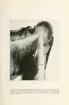
Fig. 123.
Fia. 123. Another photomicrograph from the decay shown in Figure 122, made with less ampli-
fication in order to show the relation of the decay to the pulp chamber and other parts of the tooth.
It includes a little less than one-haU of the mesio-distal section of the crown. In the preparation of
a cavity for filling in such a case, it would be an error to cut the gingival wall at d. The true cavity
lines would place the gingival wall at the line c ; the axial wall on the line b ; the occlusal wall of
the step on the line a.