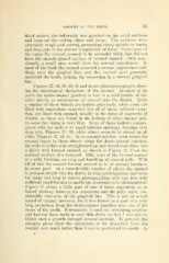
Page 119 - My FlipBook
P. 119
ATROPHY OF THE TEETH. 33
third molars, the deformity was greatest on the axial surfaces
and least on the cutting edges and cusps. The surfaces were
extremely rough and uneven, presenting sharp apiculai or knobs
and deep pits in the utmost irregularity of form. Over some of
the cusps the enamel seemed to be normally thick, but did not
have the smooth glazed surface of normal enamel. Only occa-
sionally a small area would show the normal smoothness. In
most of the teeth the enamel assumed a normal appearance sud-
denly near the gingival line, and this normal part generally
encircled the tooth, joining the cementum in a normal gingival
line.
Figures 37, 38, 39, 40, 41 and 42 are photomicrographs show-
ing the histological characters of the enamel. In most of its
parts the dento-enamel junction is lost in a wild jumble of cir-
cular whorls or protrusions of enamel into the dentin. Quite
a number of these whorls are hollow and empty, while some are
filled with amorphous material, but all of these, without excep-
tion, are lined with enamel, usually in the form of segments of
whorls, as these are found in the bottom of other enamel pits.
In some this lining is veiy thin. Some of these hollows commu-
nicate with the surface by small tubelike openings, forming very
deep pits. Figures 37, 39, while others seem to be closed on all
sides, Figures 37, 40, 42. In occasional patches, even where the
enamel began in these whorls along the dento-enamel junction,
the rods to either side straightened up and closed over them into
a fairly well formed enamel, as shown in Figure 42, from the
occlusal surface of a bicuspid. Still, most of the formed enamel
is a wild, twisting, curving and bundling of enamel rods. With
all of this the enamel formed seemed to be of normal hardness
in every part. In a considerable number of places the enamel
is plunged deeply into the dentin in long prolongations that were
too large and long to permit photographing with any lens with
sufficient amplification to enable the structure to be distinguished.
Figure 43 shows a little part of one of these appearing as an
island, midway between the cementum and the pulp canal, con-
siderably root-wise of the gingival line. This is not an actual
island of enamel, however, for it was traced as a part of a very
long projection from the dento-enamel junction near one of the
cusps of the tooth. Fortunately, I used my sectioning machine
and had cut these teeth in very thin slices, so that I was able to
follow such a growth through several sections. In general, the
pictures given show the characters of the departure from the
normal very much better than it can be portrayed in words. In