Page 101 - My FlipBook
P. 101
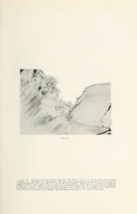
Fig. 37.
Fig. 37. Portion of buccal enamel from near the buccal cusp of a second molar from Doctor
Callow's case. Note that the den to -enam el junction, which shows plainly at the right in the picture, is
completely broken into small whorls in all of the left portion. While there is a portion of regularly
formed enamel on the right, all of the enamel on the left portion is broken or twisted out of semblance
of ordinary enamel. Also note the tubelike opening to the surface near the middle of the illustration.
These features are found to be repeated with differences of detail in the other illustrations.