Page 74 - My FlipBook
P. 74
fallen apart. These are entirely different in their mode
of growth and in size from any of the micro-organisms.
This picture is taken with the same lens as the pictures of
the micro-organisms. Dr. Noyes, who made these pictures,
has been very careful to have the comparative size exact
so that the representation on the screen and in the printed
illustrations would carry to you the idea of the difference in
size of the organisms shown correctly.
(Changing slide.) This is another species of budding
fungus, or, the Oidium Albicans. This is taken from a stale
culture where there is no mycelium shown. Some of them
are full of bright spots that represent the spores. The spores
are endogenous spores—i. e., they form inside the cell.
I have a young growth here
(Changing slides, Fig. 3.)
of the same plant. You will see something of the difference
in the form in the different periods of growth. From that
one you will see a bud starting off. Here is one nearly
separated ; here is another ; here is a young cell ; here is a
bud; there is a bud, etc. They multiply by budding, and
under certain conditions they will form a mycelium—i. e.,
'^.
^ they will form very long threads, and these will radiate in
the substance of the mucous membrane, and then they will
put up these round cells on the surface. I have seen chil-
dren's mouths white almost all over with this growth. It
produces the disease known as thrush in children.
(Changing slides, Fig. 4.) This is the next one of the
coccus series. We have here a coccus somewhat larger than
those in the first illustration. It is a streptococcus—from
streptos, a rope or a chain—that is, they hang together in
chains. Now you will notice these (pointing out) ; they seem
to be in twos; they are in the process of fission, while
these are nearly round, many of them are round. This
is the streptococcus pyogenes, one of the very ugly forms
of pus-producing micro-organisms that we find so frequently
in those very much dreaded abscesses known as carbuncles.
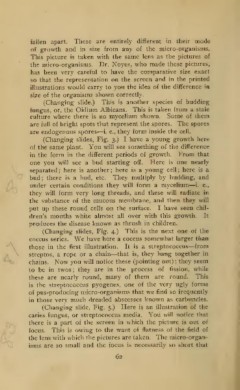
(Changing slide, Fig. 5.) Here is an illustration of the
caries fungus, or streptococcus media. You will notice that
there is a part of the screen in which the picture is out of
focus. This is owing to the want of flatness of the field of
the lens with which the pictures are taken. The micro-organ-
isms are so small and the focus is necessarily so short that
62