Page 195 - My FlipBook
P. 195
MICROSCOPICAL PHENOMENA OF DECAY 169
After the enamel has once been perforated by decay, its fur-
ther destruction proceeds principally from the inner surface.
This statement may at first seem strange, but will be found on
closer examination to be in accordance with the observed facts.
The remains of food accumulating in ever}- dental cavity do not,
of course, attack the external, but the internal surface of the
enamel. We will find, furthermore, in nearly all large cavities
the decay extending from the dentine directly upon the inner
surface of the enamel.
This latter form of decay, which we designate as secondary
enamel-decay, is in many respects better suited for study than
the primary, inasmuch as the diseased tissue is not torn away by
mastication, etc., and not contaminated from without by foreign
bodies.
The extent of the secondary decay of the enamel naturally
corresponds to that of tlie dentine-decay; in large cavities on the
grinding-surface of molars, almost the entire inner surface of
the enamel may Ije found to be involved. Penetrating enamel-
decay proceeding from within is rare. I have observed such
cases usually in inferior mo-
lars, where decay proceeding Fig. 65.
from the grinding-surface
perforates the enamel-wall
from the inner side, breaking
through the approximal wall
of enamel to the outside. In
secondary decay we find the
enamel-surface coated with a
white layer ofsoftened enamel
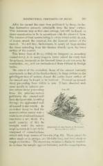
sometimes i ram. thick. If a
small quantity of this be
brought under the micro-
Disruption" of the Prisms ix Secon'dakt
scope in water, it is seen to be E.vamei.-Decay. 4'J0:1.
composed of enamel-prisms
mixed with large masses of l)acteria (Fig. 65). These prisms lie
either singly or in groups; are 10-150/i long, and have sharp or
rough extremities. The transverse striation is distinctlj' marked.
In sections the margin appt^ars indented, and the enamel-prisms