Page 96 - My FlipBook
P. 96
94 DENTAL HISTOLOGY AND OPERATIVE DENTISTRY.
the position of the cavity margins must be governed by our knowledge
of the structure of the enamel. In the execution of the work a minute
knowledge of the direction of enamel rods l)ecomes the most imjiortant
element in rapidity and success of operation.
From the standpoint of comparative anatomy, the teetli are found
to be not a part of the osseous system, but appendages of the skin,
and are to be compared with such structures in the body as the nails
and the hair. The teeth are a part of the exo-skeleton, and their rela-
tion to the bones of the endo-skeleton is entirely secondary, for the pur-
pose of strength, the bone growing up around the tooth to support it.
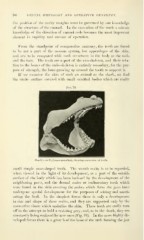
If we examine the skin of such an animal as the shark, we find
the entire surface covered with small calcified bodies Avhich are really
Fuj. 76,
Shark's skull [Lmnna cuniubira), showiiiK saeeossiuu of teeth.
small simple cone-shaped teeth. The mouth cavity is to be regarded,
when viewed in the light of its development, as a part of the outside
surface of the body which has been inclosed by the development of the
neighboring parts, and the dermal scales or rudimentary teeth which
were found in the skin covering the arches which form the jaws have
undergone special development for the purposes of seizing and masti-
cating the food. In the sim])lest forms there is only a development
in size and shape of these scales, and they are supported only by the
connective tissue which underlies the skin. These teeth are easily torn
off in the attempt to hold a resisting prey, and, as in the shark, they are
constantly being replaced by new ones (Fig. 76). In the more highly de-
veloped forms there is a growth of the bone of the arch forming the jaw