Page 68 - My FlipBook
P. 68
66 EMBRYOLOGY OF THE DENTAL TISSUES.
large polygonal cells." Dr. Williams lias shown ' that this siii)j)()si-
tion of Dr. Su(l(hith is a fact. In his photo-niicrograpiis he has
clearly demonstrated the cell contents tilling in the sj)aces between
the stellate tissue. He shows them to be very perfect nucleated cells
lying in the so-called stellate reticulum, which is reallv the sliuhtlv
modified cell wall.
The " stellate reticulum/' then, may be regarded as a storehouse of
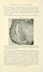
Fig. 47.
Section of jaw, cinliryo of iiiir, showing developing tooth (section teased away from tooth to show
the fold in the enamel substance): 1, enamel organ; 2, enamel substance not yet calcified;
3, layer of formed dentin ; 4, a fold in the enamel substance ; 5, dentin pulp; 6, folds at base
of dentin germ; 7, developing bone.
the calcium salts from which the first-formed layers of enamel are sup-
plied. That calcium salts exist in the meshes of the stellate reticulum
may be proven by placing a drop of dilute nitric acid on the slide when
it passes under the cover-glass. The globules or granules which were
noticed there disappear as the acid reaches them, and bubbles accumu-
late and are forced out from under the class cover. After the calcify-
,ing process commences and enamel is forming, the calcium salts are
supplied by a rich plexus of blood-vessels now in direct contact
with the cells of the stratum intermedium, all other portions of the
enamel organ having disappeared from this part. Indeed, it is difficult
to demonstrate clearly the cells of the stratum intermedium after any
' Dental Cosmos, l^ebruary, 1S96.