Page 444 - My FlipBook
P. 444
442 THE TBEATMEyT AND FILLING OF BOOT CANALS.
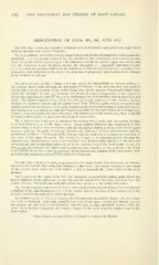
DESCRIPTION OF FIGS. 401, 402, AND 408.^
Fig. 401.—Fig. 3 gives in contrast a sectional view of deciduous and permanent upper teeth
divided tlirough their lateral diameters.
Fig. 4, a sectional view of the corresponding lower teeth divided through their antero-posterior
diameters, a, b, c represent, respectively, the deciduous and permanent front incisors in con-
trast: d, e,f, the lateral incisors ; g, h, i, the canines; k, deciduous molars, upper and lower; and
l,m, the successors to the deciduous molars, the bicuspids; n, n represent permanent molars,
c,/, i, m, o have dotted lines indicating the thickness of enamel removed by wear, atrophy of the
cementum, and reduction in the size of the pulp due to progressive calcification, these changes
being incident to old age.
Fig. 402 represents in Fig. 1, letters a to h and a to^, the longitudinal or vertical sections of
the sixteen upper teeth, showing the labio-palatal diameter of the pulp chamber and canal in
crown and roots, the section of the molars being through the anterior buccal and palatal roots,
while the bicusjiids d e and d_e illustrate the result of such a compression of the root as to
divide the pulp chamber into two canals—a condition which so frequently exists in these flattened
roots. The double-lettered series, d d to h h and d d to hh, represent in the molars a section
through the posterior buccal and the palatal roots, from whicla is quite readily recognized the
slightly greater lateral diameter of the pulp chamber in the crown and the larger canal in the poste-
rior buccal root over that in the anterior buccal root, while the bicuspids lettered eedd and ddee
illustrate a modified pulp chamber and canal, with bifurcation of the root in one, these being cut
tlirough a different axis or plane from the single-lettered series.
Fig. 2, letters a to h and a_ to h_, represent the sixteen lower teeth with the section through
their long diameters, as in the upper series. These incisors illustrate the compressed or flat-
tened condition of their roots in contrast with the cylindrical character of the roots of the upper
incisors, while the bicuspids d e and d_e illustrate the singleness of their pulp chamber and the
cylindrical condition of their roots as in contrast with the flattened or compressed condition of
the roots of the upper bicuspids. The molars /, g, h and f, g. h represent sections through the
anterior root, illustrating its compressed condition and divided pulp chamber in the first and
second molar, and a somewhat flattened one in the anterior root of the third molar ; //, g g , h h
and //, g g, h h represent the single and cylindrical pulp chamber in the posterior root of the
lower molars, while bb, ce and aa.bb represent the incisors and canines of the same series, with
modified pulp chambers arising from modified development.
Fig. 403.—Fig. 1, from a to h and a, to _^ represents the upper teeth, with transverse or horizon-
tal section through the base of the pulp chamber in the crown, viewing the entrance to the canals
of the several roots, while the same letters in Fig. 2 represent the lower series in the same
manner.
Fig. 3 represents the upper teeth, with the transverse or horizontal section made below the
largest diameter of the pulp chamber and through the canals after they have diverged from the
central chamber, but before the roots into which they run have in the molars bifurcated.
Fig. 4 in like manner represents the lower series, well illustrating the flattened or compressed
condition of the canal in anterior roots of the molars and the division of the chamber, as is fre-
quently found in the roots of the lower incisors.
The letters aa,bb,cc,d d,ff, dd. and^ (Fig. 3) represent the relative shapes, whether circu-
lar, oval, or flattened, of the pulp canal in the roots of the upper central and lateral incisors,
the canines, the first and second bicuspids, and the first, second, and third molars, while the
same letters in Fig. 4 represent the relative shapes of the pulp canal in similar teeth in the
lower series.
1 These figures are taken from v. Carabelli's Anatomie des Mundes.