Page 44 - My FlipBook
P. 44
42 MACROSCOPIC ANATOMY OF THE HUMAN TEETH.
point. The disto-lingiial ('1110:1110 is smaller than that iijxni the first
molar, and is often barely markcHl. This throws the oblique ridge more
to the distal side and enlarges the mesial fossa. The various grooves are
the same as on the first molar, except that one, the lingtial, may be lost.
The neck is less regular in outline than that of the first molar, as the
crown varies so much in shape. It is more flattened mesio-distally and
depressed toward the roots.
The roots are the same in number and general form as in the first
molar, but spread less and are more irregular in form. They may con-
verge or be crooked, or may be fused together. This makes the pulp
canals more difficult to treat. Sometimes the three roots are completely
fused, as in the third molar, and the canals may coalesce ; or the canals
of the two buccal roots may run into one. The irregularity and uncer-
tainty of the form of the roots make this tooth difficult to deal with in
treating its pulp canals.
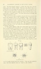
16. The Lower Molars.—The lower first molar ap])roximates the
lower second bicuspid on its distal side. It is the first of the true grind-
ers of the lower jaw and the largest tooth in the dental series. Unlike
the upper molars the transverse diameter is less than the mesio-distal.
The greater width is found across the base of the disto-buecal tubercle.
The crow'n is square or trapezoidal in form, depending on the size of the
fifth tubercle. Being quinquituberculate, the crown is broadened by the
multicuspid grinding face. The buccal face is inclined toward the centre
of the tooth, for its morsal half, to accommodate the occluding teeth.
Architecturally, the tooth is formed of four cones (Fig. 24, .4), and
Architectural diagram
(1
B
The lower flrst molar.
mav be roughly divided into four quarters. There are four primitive
cones with their tubercles and one cingule in the structure.