Page 961 - My FlipBook
P. 961
PHA GEDENIC PERICEMENTITIS. 971
ness than normal and will occasionally be deeply injected, especially if
the tooth has periods of soreness. Usually there is little or no recession
of the gum, and casual observation might not detect the presence of the
Fig. 521.
Fig. 522.
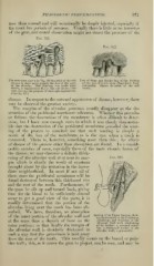
The same case shown in Fig. 519 denuded of the soft Loss of Bone and thickening of the Eorders
tissues to show more plainly the loss of the walls of the Lost Portion from Phagedenic Peri-
of the alveolus. This drawing was made after cemeutitis. Shown denuded of the soft
raising a semicircular flap ol the soft tissues over tissues,
each root for the purpose of thorough exploration.
(See Fig. 527.)
disease. In respect to the outward appearance of disease, however, there
may be observed the greatest variety.
The margins of the alveolar processes usually disappear as the de-
struction of the peridental membrane advances. Whether this precedes
or follow\s the destruction of the membrane is often difficult to deter-
mine, but I have seen enough cases in which it was clearly demonstra-
ble that the destruction of the peridental membrane preceded the wast-
ing of the process to convince me that such wasting is simply a
result of the loss of the membrane, as is the case when a tooth is
extracted. There is, however, something more than this ; for effects
of di.sea.>^e of the process other than absorption are found. In a consid-
erable number of cases, especially those of the more chronic forms of
the disea.^e, we may discover a definite thick-
ening of the alveolar wall at or near its mar- Fig. 523.
gin w^hich is clearly the result of exostosis
brought about by the irritation in the imme-
diate neighborhood. In most if not all of
these cases the peridental membrane will be
found destroyed between this thickened rim
and the root of the tooth. Furthermore,, if
the gum be slit up and turned back, giving
time for the blood to be sufficiently cleared
away to get a good view of the parts, it is
readily determined that the portion of the
alveolus lying next the tooth has been ab-
sorbed. We have, therefore, an absorption
of the inner portion of the alveolar wall and Section of an Upper Incisor show-
ing Destruction ot the Peridental
at the same time a deposit of bone on the Membrane and Eversiou of the
Alveolar Wall wiih thickening
outer portion ; so that finally the margin of of its Border: o, serumal calcu-
lus; 6, thickened border of the
the alveolar wall is decidedly thickened in alveolar wall; c, pus-cavity.
such a w^ay that the gum-tissue is held away
from the root of the tooth. . This usually occurs on the buccal or pala-
tine wall ; this, as it causes the gum to project, can be seen, and may be