Page 841 - My FlipBook
P. 841
INFLAMMATION OF THE DENTAL PULP. 851 ;
with leucocytes that have evidently escaped from the veins within a very
short time before the extraction of the tooth. This part of the section
resembles very much, except tliat the staining renders the cellular elements
far more apparent, what is seen in the early stages of inflammation in the
mesentery or web of the foot of the frog. In searching over sections of
inflamed pulps I often see little islands of inflammation in the midst
of apparently healthy tissue, as though a new nidus had been formed
at a little distance from the point of irritation or exposure of the organ.
It seems evident that these are occasionally the central points for the
formation of those minute abscesses that are so often formed in the midst
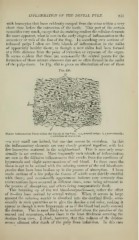
of the pulp-tissue. In Fig. 450 is given an illustration of one of these
u
Minute Inflammatory Focus within the Tissues of the Pulp: n, a, arterial twigs 6, a nerve-hundle
;
c, collection of leucocytes.
—a very small one indeed, but one that cannot be mistaken. In this
the inflammatory elements are very closely grouped together, with but
few leucocytes scattered in the neighborh(jod. This is seen only occa-
sionally in my sections. More frequently such islands of inflammation
ai'e seen in the diifusive inflammation that results from the condition of
hypersemia and slight extravasations of red blood. In these cases the
tissue is apt to be stained with the coloring matter of the red blood-cor-
puscles that have been broken up in the process of absorption. I have
made sections of a few pulps the tissues of which were thickly studded
with these ; and occasionally appearances indicate very certainly that
extravasations have occurred at different times, some being advanced in
the process of absorption, and others being comparatively fresh.
This breaking up of the red blood-corpuscles—or, rather the efl*ect
of it—has been noticed by several writers. AVhen it occurs in large
amount the coloring matter is absorbed into the dentinal fibrils, occa-
sionally in such quantities as to give the dentine a red color, making it
appear as though it were hyper^emic or as if the blood had really entered
the dentine. This is most likely to be noticed about the juncti<5n of the
enamel and cementum, where there is the least thickness covering the
dentine from view. I think, hoM'ever, that this redness of the dentine
occurs oftenest after death of the pulp from infarction. In this case