Page 697 - My FlipBook
P. 697
"
IXFLAMlLi TION. 707
boring loop, as shown in Fig. 395. These loops are formed as solid
processes, and hollowed out afterward by the removal of the central
substance of the cells. This process of the formation of ducts takes
place in the same manner in the vegetable Idngdom, and is easily
observed, especially in the sprouting of seeds. If a number of grains
of corn be planted in damp earth under suitable conditions for germi-
nation, and every twelve hours sections for microscopic examination be
made of two of these (one cut lengthwise and the other cross^vise of the
germ), the process of the formation of the ducts, which are hollowed out
in the same manner as the
blood-vessels in animals, may Fig. 397.
be followed with the greatest
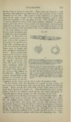
accuracy. In Fig. 397, at «,
I have represented a vow of
large cells as they appear in
the germ of the grain twelve
hours after planting, and at
b the cross-section is shown.
These grow in length, and
their number is increased by
fission. After a certain time
their growth seems to cease;
they begin to lose the central
part of their substance and
are rapidly converted into
tubes, the walls of the cells
alone remaining, which are
Formation of the Ducts in the Sprouting of a Grain of
joined together end to end. Corn, in sections cut twelve hours aftir planting. A
series of large solid cells are seen placed end to end, as
At c and d of the figure these
at 11. b is a cross-section of the same, showing the cell
are shown as they appear on to be finely granular, and staining brings the nucleus
into view. It is shown surrounded by the neighboring
the fifth day. This seems to cells, c shows the same cell converted into a tube by
hollowing out. Fifth day: conform very perfectly to the showing the elongated cells hollowed out, forming
a tube, the walls of which show " duct-uiarkiugs
manner of formation of the
(Black).
first blood-vessels in the de-
velopment of the foetus, and is much easier of accurate study.
In the animal, after the formation of vessels is once begun, all
new vessels are formed from buds given off from cells of the existing
vessels; These, though they unite A\'ith similar buds, seem to be per-
fectly fused together as a single cell. After the hollowing out is accom-
plished, however, the formed vessel presents the usual appearance of
epithelial plates joined together for the formation of its walls.
Granulation-tissue during its formation is very soft and friable. The
capillary loops come so near the surface, and their walls are so thin,
that the slightest touch is likely to cause hemorrhage, and the tissue
contains much fluid. As it grows older and the cells begin to assume
the spindle shape and form the fibrous connecting substance, it becomes
much drier and firmer. Many of the capillary loops that were formed
during the growth of the granulalions are obliterated and the tissue
shrinks, drawing the surfaces of the wounds together, usually in such a
way as to diminish its surfaces and lessen the remaining scar. This
IXFLAMlLi TION. 707
boring loop, as shown in Fig. 395. These loops are formed as solid
processes, and hollowed out afterward by the removal of the central
substance of the cells. This process of the formation of ducts takes
place in the same manner in the vegetable Idngdom, and is easily
observed, especially in the sprouting of seeds. If a number of grains
of corn be planted in damp earth under suitable conditions for germi-
nation, and every twelve hours sections for microscopic examination be
made of two of these (one cut lengthwise and the other cross^vise of the
germ), the process of the formation of the ducts, which are hollowed out
in the same manner as the
blood-vessels in animals, may Fig. 397.
be followed with the greatest
accuracy. In Fig. 397, at «,
I have represented a vow of
large cells as they appear in
the germ of the grain twelve
hours after planting, and at
b the cross-section is shown.
These grow in length, and
their number is increased by
fission. After a certain time
their growth seems to cease;
they begin to lose the central
part of their substance and
are rapidly converted into
tubes, the walls of the cells
alone remaining, which are
Formation of the Ducts in the Sprouting of a Grain of
joined together end to end. Corn, in sections cut twelve hours aftir planting. A
series of large solid cells are seen placed end to end, as
At c and d of the figure these
at 11. b is a cross-section of the same, showing the cell
are shown as they appear on to be finely granular, and staining brings the nucleus
into view. It is shown surrounded by the neighboring
the fifth day. This seems to cells, c shows the same cell converted into a tube by
hollowing out. Fifth day: conform very perfectly to the showing the elongated cells hollowed out, forming
a tube, the walls of which show " duct-uiarkiugs
manner of formation of the
(Black).
first blood-vessels in the de-
velopment of the foetus, and is much easier of accurate study.
In the animal, after the formation of vessels is once begun, all
new vessels are formed from buds given off from cells of the existing
vessels; These, though they unite A\'ith similar buds, seem to be per-
fectly fused together as a single cell. After the hollowing out is accom-
plished, however, the formed vessel presents the usual appearance of
epithelial plates joined together for the formation of its walls.
Granulation-tissue during its formation is very soft and friable. The
capillary loops come so near the surface, and their walls are so thin,
that the slightest touch is likely to cause hemorrhage, and the tissue
contains much fluid. As it grows older and the cells begin to assume
the spindle shape and form the fibrous connecting substance, it becomes
much drier and firmer. Many of the capillary loops that were formed
during the growth of the granulalions are obliterated and the tissue
shrinks, drawing the surfaces of the wounds together, usually in such a
way as to diminish its surfaces and lessen the remaining scar. This