Page 671 - My FlipBook
P. 671
VABIATIOyS IN THE BLOOD, AND IN ITS DISTRIBUTION. 681
iiig tlie contact of the escaping blood with the injured ti.ssue. When an
artery is severed, the inner coat contracts
within the outer walls— /. c. becomes the
shorter—and is pulled backward into the outer
wall of the vessel, and at the same time the
cut end is narrowed (Fig. 373). In this way
the flowing blood is brought into contact with
the greatest possible surface of injured tissue.
The formation of the thrombus is begun by
the adhesion of the wliite blood-globules to
the injured surface. These adhere one after
another, and are held fast by the formation
of a little fibrin ; others adhere, and more
fibrin is formed until the end of the vessel is
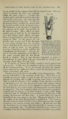
completely filled. In this process the red
globules take no part, and if the thrombus Natural H»nio.stabis. The divided
ends of the artery ((/) retract
is very slowly formed, very few of them witiiin the sheath { contractiug diminish the calibie
will be included in the clot, and it will be of the canal. IShjod coagulates in
the sheath (a) around tlie orifice
white or gray in color. In this position,
of the divided vessel, and in the
however, a thrombus is usually red from the artery itself {h) up to the first
branch (c); and lastly, plastic
entanglement of red globules. After the ar- lymph is poured out from the
divided coats of the vessel, and
tery is closed the coagulation of the blood in by its organization the perma-
nent closure takes place (Jones).
the artery proceeds until the first lateral
branch is reached. This in time becomes organized, or rather is
absorbed, and its place filled with new tissue, and the vessel is per-
manently closed (Figs. 374 and 375).
In the ligation of arteries the blood is caused to clot by injury to the
internal coat. In this case the thrombus is always red, for it contains
all the blood-constituents. If the clot should not, before the ligature
comes away, become sufficiently firm to resist the blood-pressure, sec-
ondary hemorrhage will occur.
Thrombi form in the blood-vessels under various circumstances. This
may be well studied in the mesentery of the frog. When this is ex-
posed for microscopic study, a vessel of some size may be in some way
injured—
by pricking with a needle or placing some irritant in contact
with it—and the progress of the building of the thrombus watched.
As the blood passes over the injured point a few white globules adhere
to it. Upon these others are slowly deposited in successive layers, and
the little himp is seen to grow larger and larger as the successi\'e layers
are deposited. This may continue steadily until tlie vessel is completely
occluded and the pa.ssage of the blood stopped ; or after a little clump
is formed it may be detached by some movement or by the force of the
jjassing blood-current and float away. A second clump will then be
deposited in the same manner as the first. During the growth of these
the outline of the white blood-globules is usually lost. They seem, as
the rule, to become fused with the forming fibrin into one mass, though
sometimes a few continue to show their outlines. It does not seem that
the destruction of very many white blood-globules is necessary to pro-
duce a considerable clot. The liberated material acts as a ferment, and
according to the law of the action of ferments a very little may produce