Page 66 - My FlipBook
P. 66
76 ANATOMY.
to the posterior margin of tlio otlimoidal notch, is thin and serrated,
iuid articulates with the lesser
j.j^, ,,(,
wing of the sphenoid bone.
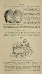
Development.—The frontal
bone is an intra-menibranous
bone, developing from two cen-
tres of ossification, which are
ileposited, one on each side above
the orbital arches, about the
seventh week of embryonal life.
At birth the bone is in tAvo por-
tions, w'hich about the first year
unite by a vertical suture (saggi-
tal) in the median line. The
Frontal Bone at Birth, developed, by two lateral halve;
union commences from below
and extends upward ; it is generally completed about the third year,
though occasionally the two halves remain sej^arate nuich later in life.
Ethmoid Bone.
The ethmoid bone (Fig. 30), like the occipital, sphenoid, frontal,
vomer, inferior maxillary, and all bones in the median line of the
skeleton, is symmetrical. It is cuboid in form, and though a true
Fig. 30.
viiA inf.'iarbinateJ b.
Ethmoid Bone, outer surface of right lateral mass (enlarged).
cranial bone it extends largely into the region of the face. It is
situated at^ the anterior part of the brain-case, between the orbits, and
fi)rms ])art of the floor of the anterior cerebral fos.'^a. "When articulated
it clo.-;cs the ethmoidal notch in the frontal bone and fi)rms the greater
part of the internal walls of the orbital cavities. It also forms the
roof and part of the septum and external walls of the nasal chambers.
It is divided into a vertical plate and lateral masses, with a horizontal