Page 641 - My FlipBook
P. 641
COMPARATIVE CHRONOLOGY OF DENTAL FOLLICLE. 651
should be felt by the practitioner, provided the teeth under his care were
erupting the upper external to the lower.
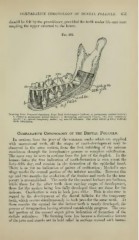
Fig. 368.
Drawing from Prepiired Supciinen from Prof. Stellwagen's Cabinet: n, permanent central incisor;
6, erupting permanent lateral incisor; c, developing permanent cuspid; Ihi, first temporary
molar; 2m, second temporary molar; ntf, mental foramen. The other letters plainly indicate
their adaptation.
Comparative Chronology op the Dental Follicle.
In sections from the jaws of the common snake which are supplied
with successional teeth, all the stages of tooth-develo})ment may be
observed in the same section, from the first infolding of the mucous
membrane through the invaginative process to complete calcification.
The same may be seen in sections from the jaw of the dogfish. In the
human foetus the first indication of tooth-formation is seen about the
forty-fifth day, and consists in the formation of the epithelial band.
There is as yet no indication of points of ossification ; ISIeckel's car-
tilage marks the central portion of the inferior maxilla. Between this
age and two months the evolution of the lamina and cords for the tem-
porary is accompli,shed. The cords for the central incisors are bulbous,
while those for the other teeth show varying stages of development,
those for the molars being less fully developed than are those for the
incisors. Ossification is seen in both jaws alike. This is also true in
regard to the development of the dental follicles for the temporary
teeth, which occurs simultaneously in both jaws for the same teeth. At
three months the enamel for the incisor teeth is nearly developed, the
process of invagination having attained considerable progress. Tlie cen-
tral portion of the enamel organ gives indication of formation of the
stellate reticulum. The forming bone has become a distinctive feature
of the jaws and stands out in bold relief in sections stained with hsemo-