Page 352 - My FlipBook
P. 352
362 DENTAL ANATOMY.
the columnar variety which still retain their connection with the Mal-
pighian layer above, from which they were orignally derived, while the
interior of the enlarged ex-
FiG- 189. tremity is composed of polyg-
onal cells.
As development proceeds, the
edges of the enlarged extremity
grow more rapidly downward
than the centre, which causes it
to assume a bell-shaped form,
with the concavity directed
downward. Synchronous with
this growth, a papilla arises
from the corium beneath and
is closely invested by the enamel
organ. The appearance of this
papilla marks the earliest stage
in the development of the den-
tine organ, but it will be well
to examine more closely at this
stage the structure of the enamel
organ. While it retained the
shape of the Florence flask its
periphery consisted of colum-
nar epithelium, the interior be-
ing made up of polygonal cells.
Coincidentally with its assump-
tion of the bell shape those cells
of the peripheral layer which are
brought into juxtaposition with
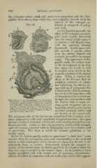
Three Stapes in the Pevelopnient of a Mammalian
Tooth-germ: o, oral epithelium heaped up over germ; the dentine bulb or organ un-
/(, younger epithelial cells; c, deep layer of cells or
rete Malpighii ; rf, inflection of epithelium for enam- dergo great elongation and en-
el germ; e\ stellate reticulum;/', dentine germ; (j,
inner portion of future tooth-sac; h, outer portion largement, forming very regular
of future tooth-sac; ?, vessels cut across; k, bone of six-sided prismatic bodies, and
jaw (from Tomes, after Frey).
are known as the enamel-cells.
The polygonal cells of the interior are transformed into a stellate retic-
ulum composed of cells with remarkably elongated processes ; these pass
through a series of unaltered cells known as the stndinn intermedium into
the enamel-cells. Lastly, we have the outer layer, which is little changed,
and still remains connected with the ^lalpighian layer by a slender cord
of epithelium. This layer is called the external epithelium of the
enamel organ.
Before the dentine papilla makes its appearance " a dark halo," more
vascular than the surrounding parts and corresponding to the epithelial
lamina or fold which gives rise to the enamel organ, is to be seen in the
submucous tissue or corium. Immediately beneath the enlarged ex-
tremity of the enamel organ the dentine papilla is developed at about the
time this stage is reac hed by the enamel organ. In its peripheral layer
highly specialized cells with several sets of processes, odontohkiHtH—
already described in connection with the tooth-pulj)—make their appear-