Page 345 - My FlipBook
P. 345
TEETH OF THE VERTEBRATA. 355 ;
mence. At the base of the crown the enamel is thrown into a conspic-
uous fokl or ridge, which completely encircles the tooth at this point, and
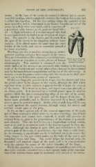
is called the cincjulum. Of the two cutting edges, the posterior is the
more extensive, and is interrupted in its descent from the summit of the
crown by a deep transverse notch, which constricts off
p i s8
a prominent cusp known as the 'posterior basal taber-
clc. A slight indication of a second cusp of this kind
is seen immediately behind it as an elevation of cingu-
lum. The anterior is the shorter, and descends from
the apex of the crown to the cingulum without inter-
ruption. It is placed nearer the inner than the outer
border of the tooth, and curves somewhat inward at
its lower extremity.
The fangs are two in number, occupying an antero-
posterior position, and give firm support to the crown.
.•', •' ' n W 1 hird Lower rreinolar
They are covered by a softer substance, resembling bone- „.,.,,
tissue, known as cementam or crusta petrosa ot human of a Dog (ranis ja-
""'"" "^' ^'^ ^"^^^^ '
This material is continued over the
odontography .
entire surface of the crown as an excessively thin stratum in the unworn
teeth of the Carnivora and several other orders, but can be demonstrated
only by the most delicate manipulation and the use of the microscope. It
assumes a more important relationship with the crown, as we shall pres-
ently see, in the herbivorous species of mammals.
Of the two fangs, the posterior is the larger, but the shorter, and takes
the greater share in the suj)port of the crown, although the cleft which
separates them at their summits is placed directly beneath the summit
of the crown. It is broad at its base, and tapers somewhat abj'uptly to
an obtuse point. It is traversed by a vertical groove upon its anterior
moiety, which fits into a corresponding ridge on the side of its socket.
The anterior root is the more slender and the longer of the two. It
tapers more gradually, and is likewise traversed by a broad, shallow
groove upon its posterior aspect. At the point of each fang will be seen
a small aperture, the apical foramen, through which the nerves and
nutrient vessels pass to the pulp.
So far, we have spoken only of the external appearance of the tooth
and of those substances which make up its outer coverings ; but if both
the cementum and enamel were removed, it would still preserve its
original form, so great is the preponderance of the dentine as a constit-
uent element. This can best be seen in a longitudinal vertical section,
since at no part in an unworn tooth is the dentine exposed in these ani-
mals. Although the dentine is quite thick, and constitutes by fiir the
greatest part of the tooth, it nevertheless does not form a solid body
on the contrary, a considerable cavity is hollowed out in its centre, this
being largest in the part which makes up the body of the crown, and
extending down each fang. This cavity lodges the dentinal pulp, the
formative and nutrient organ of the tooth, and is in communication
with the exterior by means of the apical foramina of the fangs.
While this structure, in common examples of enamel-covered teeth, is
observable with the unassisted eye, a more minute study of the organiza-
tion of the various tissues must be conducted with the aid of the micro-