Page 319 - My FlipBook
P. 319
LYMPHATIC VESSELS OF THE HEAD AND NECK. 329
spaces or alveoli are from ^ to -^ inch in diameter, and are con-
nected with each other thi'ough openings in the septa. When the
septa reach the medullary portion they subdivide and form bands or
cords which interlace in every direction and constitute a loose mesh-
work, the spaces of whieh communicate with each other and with the
alveoli. Within the meshes of the gland is contained the proper gland-
substance. In the conical compartments it is moulded into a pear-shaped
mass, while in the medullary part it assumes the form of rounded cords,
which, like the trabecular meshes, are connected with each other. In
both the cortical and medullary regions, however, there is a clear space
between the gland-pulp and the trabecule, which is termed the lymph-
)ii)ias, through wliich the lymph flows as it passes through the gland.
This lymph-sinus is crossed by a
f • TSfi
fine network of retiform connective
tissue in wliich the nuclei of the
endothelial plates covering it are
distinctly seen (Fig. 150). This
reticulation offers considerable re-
sistance to the flow of lymph
through it. The glandular sub-
stance itself consists of essentially
the same elements. It is su])port-
ed by a framework of retiform tis-
sue, in the meshes of \\hich are
found immense numbers of lymph-
corpuscles. The glandular sub-
stance is separated from the lymph-
sinus by a denser layer of reticu-
lum, although it is not so compact
as to prevent the lymph, and even
the corpuscles, from ])assing (Hit
into the lymph-sinus. The lym-
phatic glands are abundantly sup-
plied with blood-vessels. Arteries
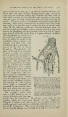
Portloii of the ISIedullary Substance of the INIes-
enter the gland at the liilum, pene- enteric filand of an Ox, tlie artery injected
with chroniate of lead (highly niagniiied) : ((,
trate into the medullary substance, medullary cylinder with cajjillary network, tine
retieuluni of connective tissue, and a tew lyniph-
and terminate in a fine capillary
corpuscles; /(, I', suijcrficial lymph-path or me-
plexus, which is surrounded and dullary sinus traversed everywhere by a retic-
ulum of nucleated cells; this reticulum has
sup])orted by the retiform tissue. been represented only at c, with numerous
anastomosing prolongations; the lymph-cor-
The veins arising from this plexus puscles have for the most part lieen removed
with a camel's hair brush ; i/, leave the gland also at the hilum. posed almost exclusively of viiistriped muscu-
The lymphatic vessels which lar tissue. A small iiied'uUary cord or liridge,
contaiuingablood-vesscl and numerous lymph-
enter a gland ramifv in the in- corpuscles, is shown at the left of the figure as
springing from the medullary cylinder.
vesting membrane, and tlieii open
directly into the lymph-sinus. The efferent vessels begin l)y fine
branches, which also communicate directly with the lym])h-sinus.
When the lymphatic vessels enter a gland they lose their external and
middle coats, and retain only the endothelial, which lines the inner sur-
face of the lymph-sinus. The current of lympli, tlierefore, can pass
directly from the afferent vessel through the lymph-sinus into the etfer-