Page 302 - My FlipBook
P. 302
312 ANATOMY.
between the posterior lacerated foramen and the entrance to the carotid
canal. From this point it ascends through the canal to the inner wall of
the tympanum, thence along a groove on the surface of the promontory,
and leaves the middle ear at its
superior and anterior portion. It
then becomes the superficial pe-
trosal nerve, and passes through
a small canal under the tensor
tympani nuiscle. Tliis canal ter-
minates in one of the small open-
iuo;s external to the hiatus Fal-
,
,% lopii. The nerve from this point
^'J extends downward through the
J petro-sphenoidal fissure or a
7 small foramen in the great wing
of the sphenoid bone, and termi-
/
nates in the otic ganglion.
Its branches of communica-
cation are two in number—one
with the carotid sympathetic
plexus, and the other witli the
tympanic plexus. After the
nerve assumes the name of the
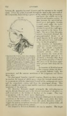
A drawing of the Tympanic Xerve (from Breschet's small superficial petrosal it is
work on the earj: A, squamous part of temporal joined by a filament either from
bone; 15, petrous portion of same; C, lower maxil-
lary nerve; D, internal carotid artery; n. tensor the geniculate ganglion of the fa-
tympani muscle; 1, carotid plexus; 2, otic ganglion
;
3, glosso-pharyngeal nerve; 4, tympanic nerve; •">, cial nerve or by the large super-
branches to carotid plexus; li, branch to fenestra
rotunda; 7, branch to fenestra ovalis; 8, branch to ficial petrosal nerve from the fa-
join the large superficial petrosal nerve; 9, small
superficial jietrosal nerve; 10, nerve to tensor tym- cial.
pani muscle; 11, facial nerve; 12, chorda tympKni;
i;^, petrous ganglion of the glosso-pharyngeal; 14, Its branches of distribution are
branch to the membrane lining the Eustachian to the tympanic plexus, the fenes-
tube.
tra rotunda, fenestra ovalis, the
promontory, and the mucous membrane of the tympanum and Eusta-
chian tube.
The pharyngeal branches (carotid branches, Henle) are three or four
in number, the largest of which passes along the internal carotid artery
to communicate with the j)haryngeal branch of the i)neumogastric nerve
and the symi)athetic system, and form the pharyngeal plexus. This
plexus snpi)lies the superior and middle constrictor muscles and mucous
membrane of the pharynx.
The Mii.scnlar Branches supply principally the stylo-pharvngeus
muscle, though filaments pass to the mucous membrane of the pharynx,
and occasionally to the borders of the base of the tongue. They may
connnunicate with the facial nerve (Rudinger).
The ToiisiUar Brandies are slender filaments which pass to the mucons-
membrane of the lower portion of the tonsillar space, where they form
a plexus (circulus tonsillaris). From this plexus branches extend to
supply the mucous membrane covering the tonsils, the palatal folds, soft
palate, and the palato-glossus muscle.
The Lingual or Terminal Branches arc two in number. The larger