Page 171 - My FlipBook
P. 171
AREOLAR TISSUE, TENDONS, AND MUSCLES. 181
Nerves.—Its supply is from branches of the inferior maxiHarv nerve.
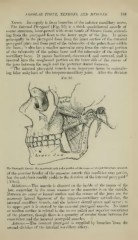
The Internal Pterygoid (Fig. 93) is a thick quadrilateral muscle of
coarse structure, interspersed with stout bands of fibrous tissue, extend-
ing- from the pterygoid fossa to the inner angle of the jaw. It arises
principally in the pterygoid fossa from the inner surface of the external
])terygoid plate and from part of the tuberosity of the palate bone within
the fossa ; it also has a smaller muscular strip from the external portion
of the tuberosity of the palate bone and the tuberosity of the superior
maxillary bone. It passes backward, downward, and outward, and is
inserted into the roughened portion on the inner side of the ramus of
the jaws between the angle and the posterior dental foramen.
" The internal pterygoid muscle is an important factor in maintain-
ing false ankylosis of the temporo-maxillary joint. After the division
Fig. 93.
The Pterygoid Muscle : the zygomatic arch and a portion of the ramus of the jaw have heen removed.
of the anterior border of the masseter muscle this condition may persist,
"
but the ankvlosis readily yields to the division of the internal pterygoid
(Allen).
Belations.—The muscle is situated on the inside of the ramus of the
jaw, somewhat in the same manner as the masseter is on the outside.
Between its outer or lateral surface and the ramus of the jaws are the
accessory lateral ligament of the temporo-maxillary articulation, the
internal maxillary vessels, and the inferior dental artery and nerve ; at
its upper part it is crossed by the external pterygoid muscle. Its inner
or median surface is related to the tensor palati and superior constrictor
of the pharynx, though there is a quantity of areolar tissue between the
constrictor and the internal pterygoid muscles.
Arteries.—The internal pterygoid is supplied by branches 'from the
second division of the internal maxillary artery.