Page 165 - My FlipBook
P. 165
AREOLAR TISSUE, TENDONS, AND MUSCLES. 175
ing from the temporal aponeurosis, the other from the mastoid process
of the temporal bone and inserted in the pinna : they are named the
attolens aurem, the attrahens aureni, and the retrahens aurem. They
are only slijjrhtly developed in man.
The AftolciiH Aurem, or Auricidaris Superior, is the largest of the three.
It is fan-shaped, arising by a bnjad head from the superfieial faseia over
the temporal muscle, and is inserted into the anterior part of the helix
and the eminence upon the inner surface of the pinna. Its fibres are
extremely delicate ; it is furnished with branches from the occipital
nerve.
The AttrdhriiH Avrcni, or Aiiricnkirin Anterior, is the smallest of the
three ; it is thin, fan-shaped, and its fibres are pale and indistinct, aris-
ing from the superficial faseia over the temporal muscle, and are inserted
into the tragus. , The nerve supplying it is derived from the facial and
the auriculo-temporal branch of the inferior maxillary.
The liefraheiis Aurem, or Avrieidaria Posterior, is stouter than the
other two, and is composed of two or three fasciculi. The •fil)res are
deeper in color and distinctly marked. It arises from the mastoid por-
tion of the temjioral bone passing forward, it narrows slightly and is
;
inserted into the posterior aspect of the concha. The nerve-supply is
derived from the posterior auricidar branch of the facial.
Muscular Action.— ^\ ith few exce])tions man has ntj power to move
the ears ; therefore the muscular action is of little or no consequence.
MUSCLES OF THE ORBIT.
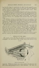
The muscles of the orbit (Figs 90 and 91) are seven in number—six
belonging to the movement of tlie eyeball : one is the elexator of the
P^iG. 90.
Muscles of the Right Orbit.
upper lid. With one exception, the seven muscles ari.«e from the back
part of the orbit, pa.ssing forward to their insertions. The other, the