Page 130 - My FlipBook
P. 130
140 ANATOMY.
Fibro- Cartilage (white) (Fig. 68) receives its name because its matrix
is composed of bundles of fibrous connective tissue, the bundles being
arranged in layers. Between the lamellae of these fibrous bundles are
rows of flattened, oval, nucleated cells, each cell being surrounded by a
delicate capsule. These cells are similar to those found in tendons, though
they are not so flat, and are distinguished by their surrounding capsule.
Where fibro-cartilage unites with tendinous tissue, as does the inter-
articulating fibro-cartilage of the temporo-maxillary articulation with
the tendon of the external pterygoid muscle, the two kinds of cells
merge imperceptibly into one another. The sesamoid cartilage, inter-
vertebral discs, interarticular cartilage, the interarticular fibro-cartilage
of the temporo-maxillary articulation, all are of the fibrous variety.
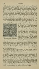
The Fibi'o-elastiG Cartilage (yellow elastic) is spoken of by some
as reticular cartilage, by reason of the
p,
arrangement of its fibres. In early
'
I ^ ; 1 life this varietv of cartilage is hyaline,
* -A: ; '. ev(^i^l;ii.' v3 but in adult life it becomes permeated
'^ with elastic fibres which proceed from
'
J
('0 the perichondrium inward. It is the
.,
•^^ -- '>^W^''^^
most elastic of all cartilage. Its fibres
are fine, and so branch and anastomose
with each other as to form a dense net-
i
Avork, with spherical or oblong spaces
'^ or meshes, in which lie nucleated cells
I
''. of varying sizes surrounded by a zone
'
,
,
,
of hyaline cartilage. This cartilage
; is
found in the epiglottis, in the auditory
'
Fibrocartilage^_of^an^Jntervertebral oauals, the Cartilage of Wrisbcrg and
Santorini, of the larynx, etc, etc.
Cartilage-cells have one characteristic which distinguishes them from
all others—viz. the power of casting around themselves a halo composed
of a substance similar to the matrix of hyaline cartilage ; this halo or
capsule, however, possesses the property of absorbing certain stains
which do not affect true hyaline cartilage. It is therefore an independ-
ent structure, and forms M'hat is known as the cartilage lacunar, ^vhich
are in reality lymph-spaces.
These lacunae are not isolated cavities, but have minute capillary
tubes communicating with each other, and finally open into larger tubes
Avhich extend to the surface of the cartilage.
Cartilage-cells are spherical or oval-shaped bodies, usually containing
one nucleus. Under certain circumstances, however, the shape of the
cell may be modified, as will be shown hereafter. They increase by
division (Fig. 70). At first the tAvo new cells formed from the original
old one are arranged side by side, in close proximity to each other, and
are half-moon shape in outline. These cells gradually separate from
each other through the increase of the capsular substance between them.
Finally, a division takes place in the capsules or lacunae and the new
cells are completed. But one cell usually occupies a lacuna, and during
the increase of cells by division each lacuna may contain two, four, six,
or eight cells, according to the rapidity of the proliferation.