Page 58 - My FlipBook
P. 58
20 PATHOLOGY OF THE HAKD TISSUES OF THE TEETH.
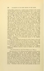
halves with a pocket lens, a curious zone of injury in the dentin
was discovered, which was photographed at once as an opaque
object, which is represented in Figure 23. Two sections, two
thousandths of an inch thick, were prepared and mounted with-
out removing them from the cover glass on which they were
ground. The sections were beautiful. No one would suspect
that there was any zone of injury in either dentin or enamel.
The disturbance of the line of the dento-enamel junction and in
the one section a clinging bit of serumal calculus were the only
abnormalities discoverable by microscopic examination. The
only way I could explain this was that the something that had
been seen and photographed had become obscured by the balsam.
The balsam was dissolved out and the section dried. A zone of
fine interglobular spaces was then found with another singular
appearance in the form of a broad line of demarkation, that could
not be explained. The section was remounted in a very stiff
balsam without using anything to clear the dentin, with the
expectation of making a photomicrograph the same evening.
Something prevented, and by the next evening, the day having
been unusually warm, the interglobular spaces were again filled
with balsam. The shadow, however, remained, and is presented
in Figure 24. It has since been found that the condition pre-
sented is common to a considerable number of the slighter
injuries of this type.
Figure 25 is a photomicrograph of a labio-lingual section cut
from near the mesial, side of a malformed tooth so that the line
of interglobular spaces is cut through diagonally. This gives
an exaggerated view of the zone of injury to the dentin, but will
serve to impress the fact that these injuries are very severe.
This presents this subject from its gravest to its slightest
degree, in sufficient variety of cases to render the conditions
intelligible.
The Deformity in the Fiest Permanent Molars.
The deformity of the first permanent molars should receive
special consideration because of its greater frequence and
because it so generally leads to early and rapid caries beginning
in the malformed portion. The plan of injury does not differ
from similar deformities in the front teeth, but the details of the
injury are different because of the wide difference in the form of
the tooth. Greater frequence of the occurrence of the condition
in these teeth is due to the earlier beginning of calcification. In
dissections of the jaws of the fetus at term, I have usually found
the calcification of this tooth just begun on the points of the
cusps. Sometimes there are only small spicuke, in other cases