Page 274 - My FlipBook
P. 274
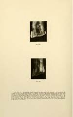
Figs. 160, 161. Photographs of split incisors that have been badly abraided. In Figure 160 the
outlines of what was the pulp chamber are sharply apparent in the incisal portion, but the whole pulp
chamber is solidly filled with a calcific deposit which has obliterated the dentinal tubules, cutting off
all connection of the dentinal fibrils with the pulp of the tooth. This has destroyed the vitality of the
crown of the tooth and obliterated all sensation in the dentin. In Figure 161 there is very nearly the
same condition, but in the light line in the central portion of the pulp canal there was a shred of
living pulp tissue that may, or may not, have retained connection with a few of the dentinal fibrils
of the crown of the tooth.