Page 142 - My FlipBook
P. 142
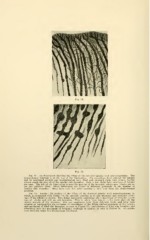
Fig. 74.
Fig. 73. An illustration showing the filling of the dentinal tubules with mi iroorganisms. The
dento-enamel junction is at the top of the illustration. The organisms have entered the tubules
and by continued growth and multiplication have filled and enlarged them i ery evenly, having
grown single file into all of the smaller side branches. Only a few slight local swellings of tubules
are seen. The left of the illustration is near the margin of the invasion, where new tubules are as
yet but partially filled. Great differences are found in different specimens in the number of
smaller side branches. Some have very few after passing a little way fror i the dento-enamel
junction.
Fig. 74. Another illustration of the filling of the dentinal tubules with lcroorganisms, in
which, as compared with Figure 73, the opposite extreme as to side branches ; d irregular swell-
ings of the tubules figure represents something like >f irregular swell-
ings of tubules and with n hes. Th deeper in the tooth close on the
deeper margin of the invi two specimens i different teeth, and have been
selected as representing the extremes of sm< of the filling and enlargement of the tubules
and maximum of filled side branches, as seen n Figure 73 ; and absence of filled side branches and
something near the maximum of irregular s ellings of tubules, as seen in Figure 74. All grada-
tions between these two illustrations are fou