Page 612 - My FlipBook
P. 612
296 THE TECHNICAX, PROCEDURES IN FILLING TEETH.
food gathering between the teeth at meal time, and the discom-
fort and pain this produces. By so forming the contact and
contour of the proximal surface that there will be very free
excursions of food through the emljrasures in chewing, the mar-
gins of fillings will be well cleaned and the danger of recurrence
of decay greatly limited. All of these considerations call for
close clinical study of the forms of proximal surfaces best suited
to these ends.
Form op contact point. The form of the immediate jDoint
of contact should be similar to the rounding of the surfaces of
two marbles when placed in contact, and if this rounding can be
continued for a space toward the gingival and then the curve
reflected to form a concavity, followed by a straight line toward
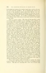
the gingival, as shown diagrammatically in Figures 397, 398,
the form is improved for the jjurposes intended, i. e., the pre-
vention of food lodgments. In this case any stringj" food
particles that are forced past the contact point will be loose
the moment the contact point is passed and will be pulled away
by the next crush that forces food through the embrasures.
Figures 396, 397, 398, show the outlines of contact points and
proximal surfaces found between the second bicuspid and first
molar. Figure 396 may be regarded as a fairly good form, for
it lias a reasonably wide interproximal space at the gingival line.
Figure 397 is better, not so much because of the increased
breadth of the interproximal space at the gingival as by the
increased curve at the contact point and the reflection of the
curve producing a concavity in the line toward the gingival.
Therefore, Figure 397 is a better form than Figure 396. The
form represented in Figure 398 is still better in the harmony of
its lines, and represents about the extreme of relative breadth
of teeth and interproximal space found normally in strongly
bell-crowned teeth. The rounding from the occlusal surface to
the point of contact is much better in Figures 396 and 398 than
in Figure 397, and will serve a better purpose. With such forms
as represented in Figures 397, 398, the interproximal gum tissue
will continue to fill the space almost completely to a good old
age, if not injured by deposits of calculus. Soft tissue filling an
interproximal space terminating in a point so acute as that
shown in Figure 396 is not so well maintained and tends to
shorten, leaving the space open much earlier.
These observations make it desirable to imitate the forms
given in Figures 397, 398, so far as possible in forming proximal
surfaces. The files for trimming these have been formed with