Page 425 - My FlipBook
P. 425
^
Fig. 20!).
Fig. 268.
Fig. 27 u.
L.
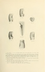
Figs. 265-271. A series representing extensive caries in the mesial surface of an incisor tooth
and its treatment.
Fig. 2fio. An incisor tooth with extensive decay in the mesial surface showing a wide-open
cavity. It happens sometimes that when the enamel has broken so as to open the cavity very wide
early, tliat the rapidity of penetration has been checked while the decay has continued to extend along
the dento-enamel junction, making extensive undermining before exposure of the pulp occurs.
Fig. 266. A preparation of the cavity retaining as much enamel as practicable on the labial
surface and cutting around the pulp to prevent its exposure.
FjG. 267. A cross section of the prepared cavity close to iho gingival wall, showing the plan
of obtaining anchorage.
Fig. 268. The cavity as prepared after removal of the pulp.
Fig. 269. The cutting of the pulp canal in order to obtain the straighte.st path for the broach
in removing the pulp from the canal and filling the root.
Fig. 270. The completed filling after eithir mode of preparation.
Fig. 271. The filling from the labial after either mode of preparation.