Page 399 - My FlipBook
P. 399
Fig. 23!).
Fig. 243.
Fig. 244.
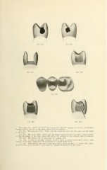
Figs. 239, 240. Distal and mesial views of the first bicuspid, showing the decays. Examination
reveals extensiTc exposure of the pulp, which must be removed.
Fig. 241. The prepared cavity. Notice that the enamel has been cut away just over the points
of the cusps.
Fig. 242. The cavity filled. Notice that the filling material protects the entire occlusal surface
so that the danger of the cusps being split off by the wedging of food between them is removed.
FiQ. 243. The finished case as seen from the occlusal view.
Fig. 244. A view of the first bicuspid, as often filled, in mesio-occlusal-distal cavities after
removal of the pulp, which gives the appearance of a much better tooth.
FiQ. 245. This exhibits the usual result of a filling placed as shown in Figure 244, which,
sooner or later, is pretty sure to occur from the wedging of food between the cusps.