Page 239 - My FlipBook
P. 239
THE HISTOLOGICAL STRUCTURE OF THE TEETH. 99
cavity wall is most generally so close to the angle as to be out
of the way. Yet, it should always have careful attention. Cavi-
ties in the mesial surfaces of the upjaer incisors can therefore
approach the incisal angle more closely with safety and afford
opportunity for a good margin for a filling — without too thin
an edge, than can cavities in the distal surfaces of these teeth.
In following the inclination of the enamel rods around the
incisors and cuspids in the circumferential direction, we find
them generally standing peri)endicular to the surface at the junc-
tion of the middle and gingival third of the length of the crown.
A notable exception to this, is the approach to and over the
mesio- and disto-liugual marginal ridges. Here the enamel rods
incline somewhat toward the marginal ridges, but, in passing
over these ridges, their direction or inclination changes very
suddenly and often very irregularly. For this reason this
becomes rather a dangerous point in the preparation of proximal
cavities in the incisors. When the marginal lines of these proxi-
mal cavities reach to the lingual marginal ridge, it is rarely safe
to leave any of the ridge remaining, because of the very uncer-
tain direction of the enamel rods. Especially is this true of
lateral incisors in which the curve of the ridge is often very
abrupt. While this I'idge is very strong in the perfect tooth, it
becomes very frail when its support on either side has been
destroyed, and the only safe course seems to be to cut it away
sufficiently to be certain of the direction of the enamel rods upon
the margin formed. The rounding of the labio-mesial or labio-
distal angles is not so abrupt, and the enamel rods usually hold
closely to a direction perpendicular to the surface, so that good
margins can be made at any point by observing carefully the
form of the tooth and the enamel cleavage.
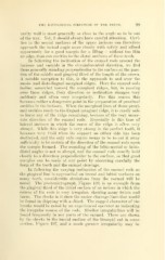
In following the varying inclination of the enamel rods as
the gingival line is approached on buccal and labial surfaces on
many teeth, considerable deviations from the normal will be
noted. The photomicrograph, Figure 109, is an example from
the gingival third of the labial surface of an incisor in which the
course of the rods is very irregular, showing many twists and
turns. The checks in it show the easier cleavage lines that would
be found in chipping with a chisel. The ragged character of the
breaks would be noted by an experienced operator as indicating
the irregular course of the rods. Similar irregularities will be
found frequently in any parts of the enamel. These are shown
by the checks in the buccal surface of the bicuspid cut in cross
section, Figure 197, and a much greater irregularity may be